A new imaging system may help clinicians to identify early bone damage in the hands of people with rheumatoid arthritis (RA) at a time when vast improvements are still possible.
![]()
Research has shown that people with RA (who are more commonly women aged between 25 and 50) are at an increased risk of developing permanent bone damage. Most often, it’s in the hand bones that this damage first occurs, causing disfigurement and profound loss of hand functioning. Disease modifying anti-rheumatic drugs (DMARDs) have proven to be an effective treatment for limiting the damage before it severely debilitates. Yet, if the bone damage goes unrecognized and progresses too far, it becomes too late for these drugs to work.
How can clinicians be sure to identify bone damage early enough to halt further bone thinning and erosion?
To date, clinicians have had to rely mainly on patients’ reports of their symptoms. Imaging
technologies such as X-rays and MRIs have only been able to identify macro-structural bone changes that become apparent once bone damage is already irreversible.
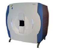
The new imaging system – high-resolution peripheral quantitative computed tomography (HR-pQCT) – could aid clinicians’ ability to diagnose. Images of the finest details in bone (often so fine they are similar in thickness to a human hair) can be captured by this new technology. As a result, scientists are hoping that HR-pQCT can identify micro-structural bone changes prior to permanent damage occurring in people with RA.

To test the potential of this new technology, Dr. Lynne Feehan’s team are imaging the hand and forearm bones of 30 participants who have been diagnosed with RA in the last 12 months. Images are also being taken from a comparison group of participants without inflammatory arthritis, matched for age and sex. All participants will be followed for one year to see if HR-pQCT can reliably identify and track micro-structural changes in bone over 12 months. This new technology may lead to better monitoring of early disease progression and the effects of RA treatments on bone health.
Principal Investigators:
Heather McKay
Linda Li
Lynne Feehan
Co-Investigator:
Kam Shojania
Collaborators:
Steve Boyd
Helen Buie
Cheryl Barnabe
Very nice style and excellent articles, nothing at all else we need :D.
Hello. remarkable job. I did not anticipate this. This is a fantastic story. Thanks!
I think you have remarked some very interesting details , regards for the post.
I was examining some of your content on this website and I think this web site is rattling informative! Keep putting up.